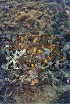
Masson’s trichrome blue (MTB) staining was used to demonstrate the quantity of fibrillar collagens in HMD and LMD tissue sections, and Ki-67 antibody (clone MIB-1, catalogue number M7240 Dako, Carpinteria, CA, USA) was used for assessing and comparing proliferation status. Immunohistochemical Masson’s trichrome blue stainingĪ total of 15 pairs of HMD and LMD tissue samples were selected, based on the presence of epithelium on H&E-stained images, to represent the characteristics of the mammary specimens. The tissue samples were then fixed in 10 % neutral buffered formalin, processed through alcohol, embedded in paraffin blocks and then sectioned at 5-μm thickness for histological analyses.

This method was detailed in our previous work. When the tissue slice mammogram appeared homogeneously grey or lacked black-white differential, the patient was excluded from the study. The tissue slice was transferred to a biosafety level 2 hood, where areas that appeared white on X-rays were selected and removed using sterile blades and defined as HMD regions, and areas that appeared black were similarly selected, removed and classified as LMD regions. X-rays of those slices were taken by a breast radiographer using uniform radiological parameters and were assessed against a calibration ruler for selection of HMD and LMD regions. Resected breast tissue was transferred immediately to the pathology department upon completion of mastectomy, where pathologists resected slices of breast tissue using a sterile technique. Tissue accrual and selection of high and low mammographically dense regions In an effort to assess the possible role of epithelial–mesenchymal transition (EMT), which has been implicated in many aspects of malignancy, we included vimentin in our analysis of epithelial cells. We assessed differentiation, proliferation, hormone receptor and aromatase expression in the epithelium collagen content and organisation and immune cell status. In an attempt to define the tissue components responsible for the increased MD, we analysed the epithelial, stromal and adipose compositions and further investigated the stromal and epithelial compartments. The abundance and phenotype of immune cells, now an acknowledged component of the tumour microenvironment, have also never been assessed in HMD tissue. The composition of the stroma has been examined in a few studies, which have shown that the degree of collagen deposition is increased in HMD tissue however, collagen organisation has not been assessed.

The proportion of basal and luminal cell layers within the epithelium has never been assessed, and change in this proportion is one of the first steps in tumorigenesis. HMD tissue has been shown to be composed of greater areas of stroma, but less fat, than LMD tissue however, findings on whether HMD area contains higher amounts of epithelium are conflicting, and epithelial cell proliferation appeared to be increased in some studies, but not in others. To date, various studies have examined the histological and molecular differences of HMD and low mammographic density (LMD) breast tissues. As the biological basis of MD is not understood, viable preventative and/or therapeutic targets aimed at reducing MD-associated BC risk have yet to be developed. As of December 2014, Breast Density Inform laws, which call for mandatory reporting of MD status, had been introduced in 20 states across the United States. Advocacy groups such as Are You Dense? have provided education to the public and helped advocate changes in public policy. The link between high MD and BC risk is now in the public domain. By contrast, the prevalence of HMD suggests that as many as 30 % of BC cases could be attributable to elevated MD, should the MD–BC risk association be causal. Although 55 % to 65 % of BRCA1 mutation carriers develop BC by the age of 70, these cases are estimated to account for just 2 % to 5 % of all BCs. In a study of 1353 women in America, researchers found that 42 % of the 40- to 59-year-old age group and 25 % of the 60- to 79-year old age group have breasts that are at least 50 % mammographically dense.


High mammographic density (HMD) is not uncommon in the community. Women with breasts that have 75 % or greater mammographically dense tissue are four to six times more likely to develop BC than are women with breasts that have 10 % or less mammographically dense tissue, and this risk has been shown to persist over 10 years of follow-up. The radiological appearance of the breast differs among women owing to varying proportions of radiodense fibroglandular tissue that appears white and radiolucent adipose tissue that appears dark. Important risk factors for breast cancer (BC) are increasing age, genetic mutations, family history or personal history of BC and mammographic density (MD), which refers to the extent of opaque appearance on a mammogram.


 0 kommentar(er)
